Case 2: Dose Management in Endovascular Image-Guided Neuro-Interventions
CE and CME currently unavailable
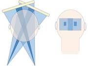
Describes the effective use of collimation and region of interest imaging to reduce dose during fluoroscopy.
Authors: D.R. Bednarek; C. Ionita; S.N. Sweadri Vasan; S. Rudin
Target Audience: Radiologists, radiologic technologists and medical physicists working with fluoroscopy
This educational activity is free.
Learning Objectives
Upon completion of this case, the participant will be able to:
- Recognize the benefit and importance of x-ray beam collimation for reducing stochastic risk
- Utilize region of interest (ROI) imaging and demonstrate how it can be used to reduce integral dose
- Use collimation and ROI imaging to reduce peak skin dose